As an applied scientific discipline, the study of Chinese medicine can involve:
- Identifying the medicinal ingredients
- Validating the quality
- Developing quality standards
- Finding and exploring new sources of medicinal ingredients
Although traditional Chinese medicine has been practiced for thousands of years, today this discipline incorporates modern scientific methods, including microscopy.
Methodology Used to Identify Chinese Medicinal Ingredients
Samples used to identify and validate Chinese medicinal ingredients can be in several forms, including intact pieces, fragments, tablets, and powders. The different sample presentations require diverse identification and validation methods.
Four methods are commonly used:
- Source investigation
- Trait characterization
- Microscopy examination
- Physical and chemical analysis
Often, the inspector needs to use multiple methods to identify and validate a sample according to its condition and identification and validation requirements.
The microscopy inspection method involves using a microscope to observe an ingredient’s tissue structure and cell shape, as well as the characteristics of its contents. Usually used to assess an ingredient’s authenticity and purity, this method is also commonly used when it’s difficult to identify a sample based on its exterior physical condition or if the sample has been broken down into small pieces or a powder.
Digital Microscopy: Macroscopic to Microscopic Observations Made Easy
For ingredients in the form of chopped herbs and leaves, the inspector typically prepares a slide with a surface slice to observe the microscopic details. When identifying and validating animal-based medicinal materials, macroscopic observation of the whole sample is particularly important.
With a magnification range of 20x to 7000x, the Olympus DSX1000 digital microscope enables inspectors to perform high-quality observations of whole specimens at low magnification and then quickly switch to micron-level magnification for a detailed analysis. It also offers a large depth of field and a long working distance, so inspectors can examine large samples. And, using the DSX1000 microscope's tilt model, inspectors can capture images of samples from multiple angles.
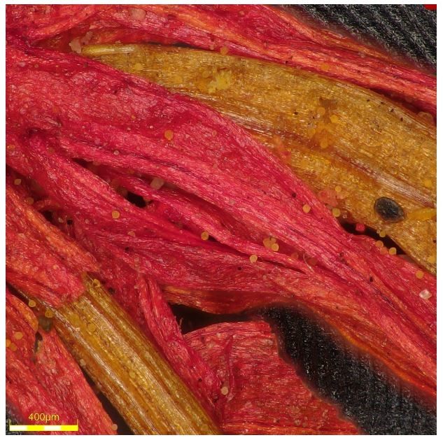
Histomorphology of saffron magnified to 400 µm
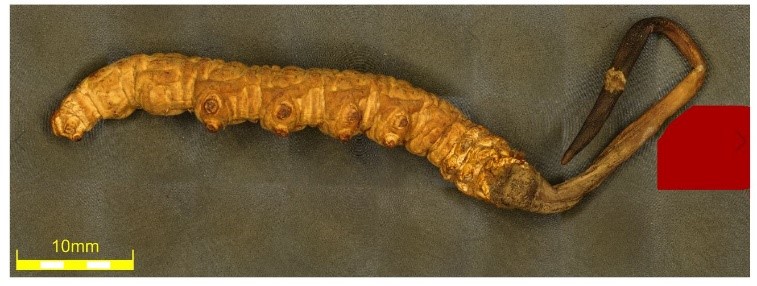
Caterpillar fungus (Ophiocordyceps sinensis) macro image
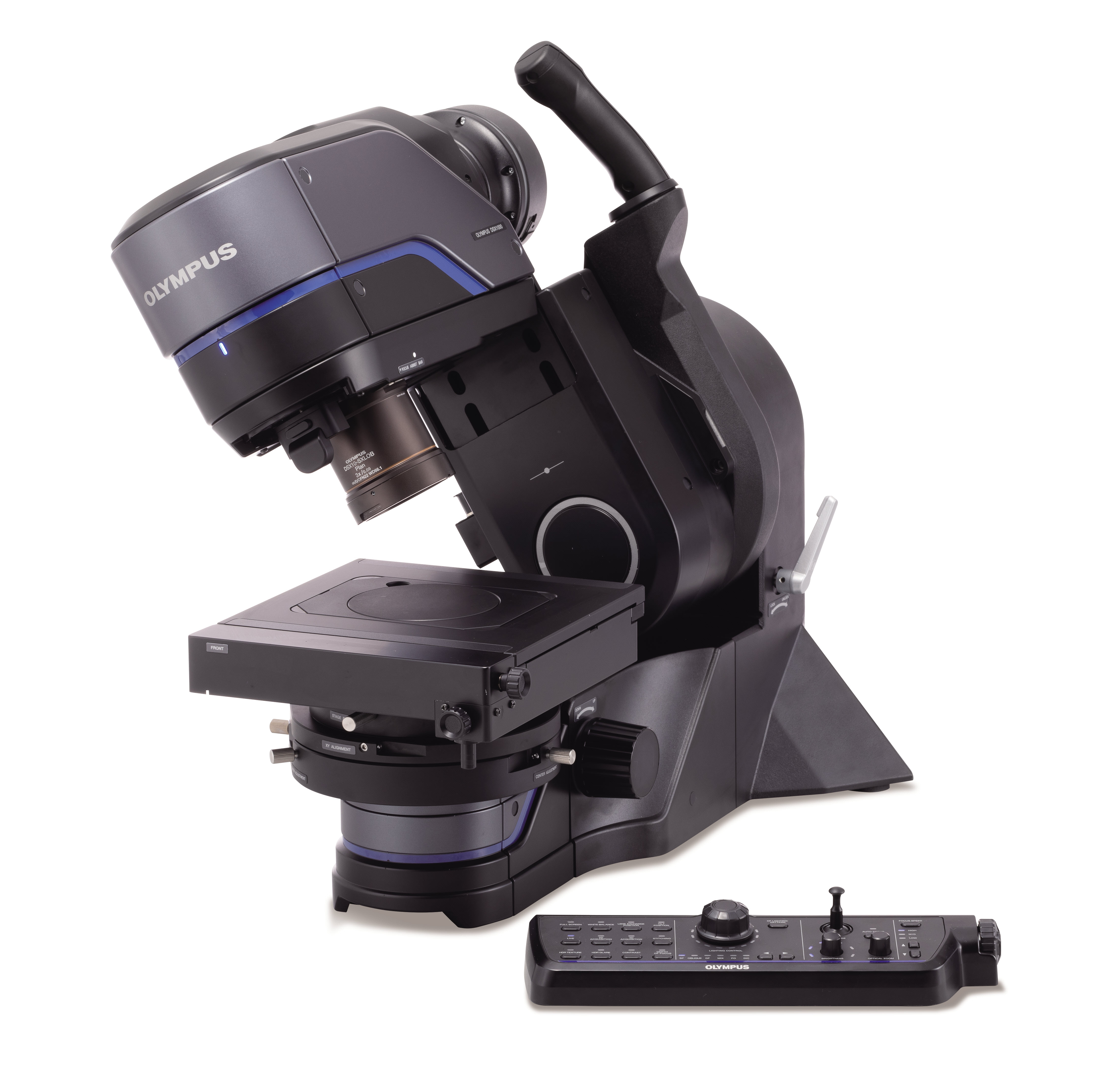
DSX1000 digital microscope tilt model with its remote-control console
Simplified Selection between Six Observation Modes
Detecting traits of certain plant-based ingredients can be facilitated by polarized light observation. This is because their tissues, cells, and contents display consistent polarization phenomena. For instance, the various starch granules of the astragalus plant (an herb with purported immune-boosting, anti-aging, and anti-inflammatory effects) and the calcium oxalate crystals in ginseng can be detected using polarized light.
Inspectors can also use the microscope’s polarized light observation mode on finely ground or microsectioned mineral samples to quickly and accurately detect polarization traits. For example, in transparent or translucent minerals, the inspector can examine and validate thin-slice samples by observing the refraction, reflection, and interference behavior of the light that enters the mineral’s crystal
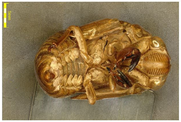
Macro imaging of a cicada molt sample
A conventional microscope that only offers one or two observation methods provides limited information from the sample. The DSX1000 digital microscope features multiple observation modes. Moreover, it has a “Best Image” function that makes it simple to choose the optimal observation mode.
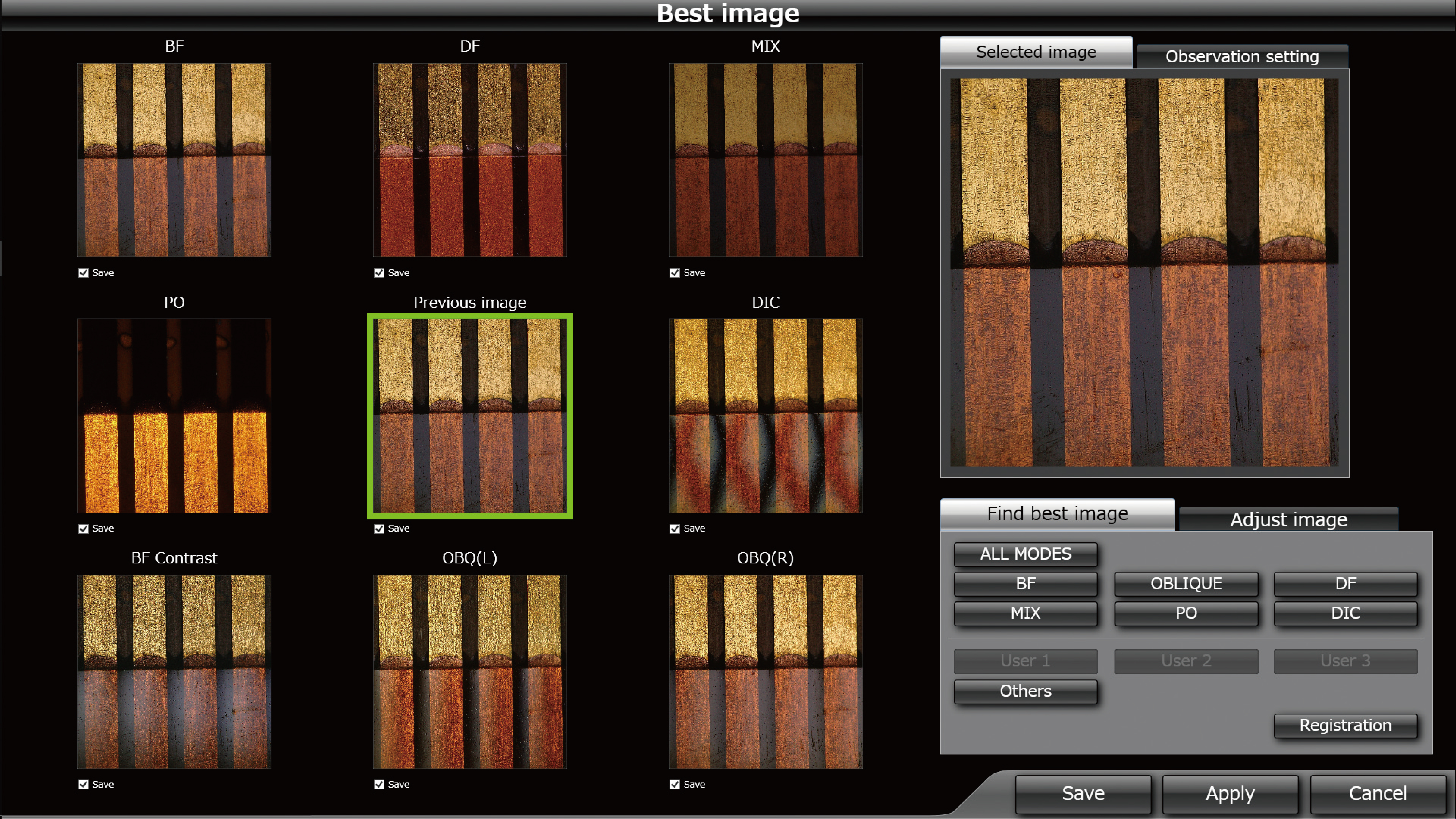
The microscope's Best Image function enables you to compare and select the optimal observation mode
Images of the six observation modes are displayed on the screen, BF (brightfield), OBQ (oblique), DF (darkfield), MIX (brightfield + darkfield), PO (simple polarization), and DIC (differential interference). The inspector just taps their preferred image to select the mode.
To learn more about the advanced features of the DSX1000 digital microscope, visit www.olympus-ims.com/microscope/dsx/ or contact your local Olympus representative.
Related Content
5 Advantages of the DSX1000 Digital Microscope
From Macro to Micro: How Digital Microscopes are Changing Industrial Inspections
Discovering the Unseen Beauty of Sand Under a Digital Microscope
Get In Touch
