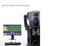
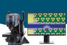
Overview
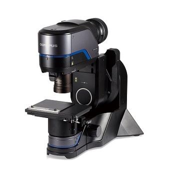
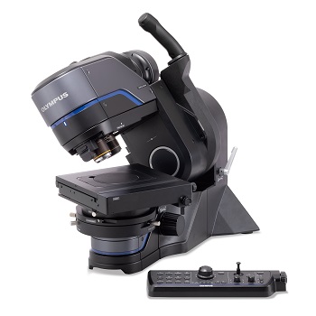
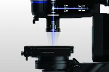
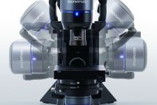
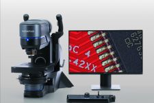
See Your Sample from Many AnglesDSX1000 Tilt Model |  |
|---|
See the Whole Picture:
|
|---|
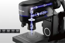
 | Instant Switching Saves Time
|
|---|
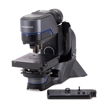
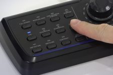
Easy-to-Use Console
*To guarantee XY accuracy, calibration work must be undertaken by an Evident service technician. | 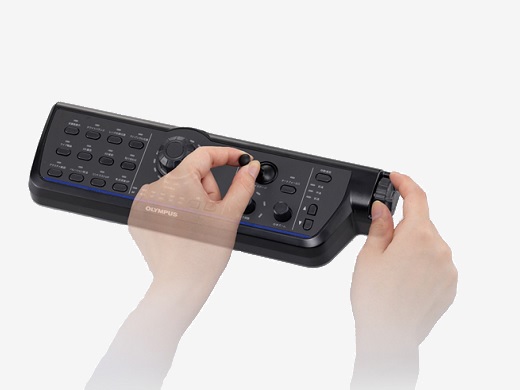 |
|---|
5 Advantages of the DSX1000 Series Digital Microscope Over Conventional Digital Microscopes
|
Back to Digital Microscopes
Models
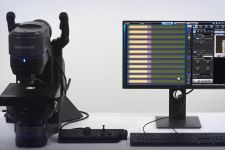
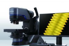
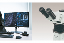
DSX1000 Digital Microscope LineupYou can meet various observation needs with the DSX1000 Series from entry level to high end.
|
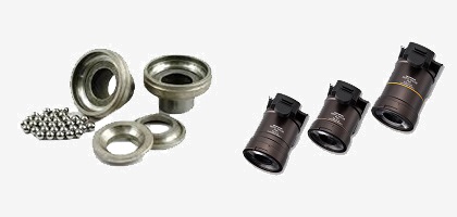
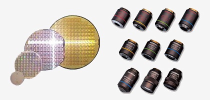
DSX1000 Objective LensesOur lineup of 17 objective lenses, including super long working distance and high numerical aperture options, offers flexibility to obtain a wide range of images.
|
Back to Digital Microscopes
Specifications
DSX1000 Digital Microscope Specifications |
| DSX10-SZH | DSX10-UZH | |||
| Optical System | Optical system | Telecentric optical system | ||
| Zoom ratio | 10X (motorized) | |||
| Zoom magnification method | Motorized | |||
| Calibration | Automatic | |||
| Lens attachment | Quick-switch, coded lens attachments automatically update magnification and visual field information | |||
|
Maximum total magnification
(on a 27-inch monitor, 1:1 display, at 100% image magnification) | 9637X | |||
| Working distance (W.D.) | 66.1 mm – 0.35 mm | |||
| Accuracy and repeatability (X-Y plane) | Accuracy*1 | ± 3% | ||
| Repeatability 3σn-1 | 2% | |||
| Repeatability (Z axis)*2 | Repeatabilty σn-1 | 1 μm | ||
| Camera | Image sensor | 1 / 1.2 inch, 2.35 million pixel color CMOS | ||
| Cooling | Peltier cooling | |||
| Frame rate | 60 fps (maximum) | |||
| Low | 960 × 600 (16:10) | |||
| Medium | 1600 × 1200 (4:3) / 1920 × 1080 (16:9) / 1920 × 1200 (16:10) / 1200 × 1200 (1:1) | |||
| High (pixel shift mode) | 2880 × 1800 (16:10) | |||
| Super high (pixel shift mode) | 5760 × 3600 (16:10) | |||
| 3CMOS mode (high quality) | Not available | Available (high and super high modes only) | ||
| Illumination | Color light source | LED | ||
| Lifetime | 60,000 h (design value) | |||
| Observation | BF (brightfield) | Standard | ||
| OBQ (oblique) | Standard | |||
| DF (darkfield) |
Standard
LED ring divided into four divisions | |||
| MIX (brightfield+darkfield) |
Standard
Simultaneous observation of BF + DF | |||
| PO (polarization) | Standard | |||
| DIC (differential interference) | Not available | Standard | ||
| Contrast up | Standard | |||
| Depth of focus up function | Not available | Standard | ||
| Transmitted lighting | Standard*3 | |||
| Focus | Focusing | Motorized | ||
| Stroke | 101 mm (motorized) | |||
*1 Calibration by an Evident or a dealer service technician is necessary. To guarantee the accuracy of XY, calibration with DSX-CALS-HR (calibration sample) is required. To issue certificates, calibration work must be undertaken by an Evident calibration service technician.
*2 When using a 20X or higher objective.
*3 The optional DSX10-ILT is required.
| Objective | DSX10-SXLOB | DSX10-XLOB | UIS2 | |
| Objective lens | Maximum sample height | 50 mm | 115 mm | 145 mm |
|
Maximum sample height
(free angle observation) | 50 mm | |||
| Parfocal distance | 140 mm | 75 mm | 45 mm | |
| Lens attachment | Integrated with lens | Available | ||
|
Total magnification
(on a 27-inch monitor, 1:1 display, at 100% image magnification) | 27 – 1927X | 58 – 7710X | 34*4 – 9637X | |
| Actual F.O.V. (μm) | 19,200 µm – 270 µm | 9,100 µm – 70 µm | 17,100 µm – 50 µm | |
| Adaptor | Diffusion adaptor (optional) | Available | Not Available | |
| Eliminate reflection adaptor (optional) | Available | Not Available | ||
| Lens attachment | Number of objectives that can be attached |
Up to 1 piece
(attachment is integrated with lens) | Up to 2 pieces | |
| Objective lens case | Three lens attachments can be stored | |||
*4 Total (maximum) magnification when using MPLFLN1.25X
| Stage | DSX10-RMTS | DSX10-MTS | U-SIC4R2 |
| XY stage: motorized / manual | Motorized (with rotation function) | Motorized | Manual |
| XY stroke |
Stroke priority mode: 100 mm × 100 mm
Rotation priority mode: 50 mm × 50 mm | 100 mm × 100 mm | 100 mm × 105 mm |
| Rotation angle |
Stroke priority mode : ±20°
Rotation priority mode : ±90° | Not available | |
| Display rotation angle | GUI | Not available | |
| Load-resistance | 5 kg (11 lb) | 1 kg (2.2 lb) | |
| Frame | DSX-UF | DSX-TF |
| Z-axis stroke | 50 mm (manual) | |
| Tilt observation | Not available | ±90° |
| Tilt angle display | Not available | GUI |
| Tilt angle method | Not available | Manual, fix / release handle |
| Measurement | Standard | Basic interactive measurements |
| 3D Line profile measurement and simple 3D measurements | ||
| 2D Line profile measurements | ||
| Advanced interactive measurement, including auto-edge detection and auxiliary lines | ||
| Neural Network Labelling | ||
| Live AI | ||
| Offline EFI, Offline Panorama | ||
| Image enhancement filters | ||
| Optional | 3D Analysis Application* | |
| Count and Measure | ||
| Neural Network Training | ||
| Material Solutions | ||
| Auto edge measurement | ||
| Particle analysis | ||
| Sphere/cylinder surface angle analysis | ||
| Multi-data analysis** |
*Requires PV-3DAA.
**Requires Experimental total assist application software (OLS51-S-ETA).
| Display | 27-inch flat panel display |
| Resolution | 1920 (H) × 1080 (V) |
| System total | Upright frame system | Tilt frame system |
| Weight (frame, head, motorized stage, display, and console) | 43.7 kg (96.3 lb) | 46.7 kg (103 lb) |
| Power consumption | 100 – 120 V / 220 – 240 V, 1.1 / 0 .54 A, 50 / 60 Hz | |
Back to Digital Microscopes
Applications
DSX1000 Applications |
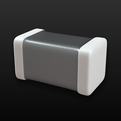 | Perform Highly Accurate Thickness Measurements of the Internal Layer of a Multilayer Ceramic CondenserMultilayer ceramic condensers (MCLLs) have been attracting attention and it has found widespread use in applications ranging from mobile terminals to automobiles. Moreover, it is expected that large quantities of MCLLs will be incorporated into 5G devices. The DSX1000 it easy to measure thickness of the Internal layer of MCLLs with high resolution. |
|
|---|
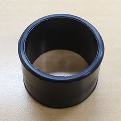 | Using a Digital Microscope for Precise Burr Measurement on Injection-Molded ProductsOlympus' DSX1000 digital microscope makes it easier to obtain optimal images that facilitate the quality control of burrs on injection-molded components. It comes equipped with various functions that enable you to acquire images at the desired magnification, observation method, and illumination angle, and an image-processing function. |
|
|---|
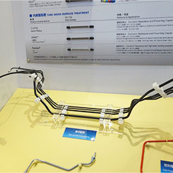 | Measuring the Thickness of Automotive Pipe Coatings Using a Digital MicroscopeIn the quality control process, inspectors must assess coating thicknesses to make sure they meet specifications and check for thickness variations. DSX1000 provides pattern matching and shading correction algorithms that enable you to stitch images together. |
|
|---|
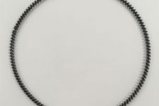
 | Inspecting Burrs on Pistons Using a Digital MicroscopeIf there are burrs in the piston’s grooves, it can lead to serious engine issues. DSX1000 offers "Observe small burrs with clear images at low magnification" , "Instantly switch to a higher magnification objective to analyze burrs" and " See the piston ring groove from different angles with a tilting frame" and provide efficient workflow. |
|
|---|
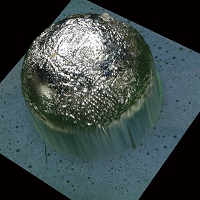
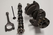
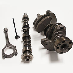 | Observing the Metal Flow in Forged Products Using a Digital MicroscopeThere are many parts that are forged, such as gears, valves, and connecting rods used in automobiles. DSX1000 can observe the metal flow that affects toughness using the auto-stitching function. |
|
|---|
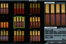
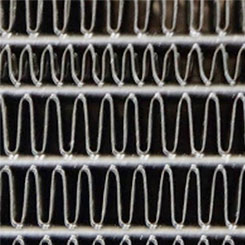 | Inspecting the Brazed Joints of Radiator Fins Using a Digital MicroscopeRadiator are important role in engine cooling and it is essential to confirm the brazing of pipes and fins for quality control. DSX1000's multi-preview function makes it simple to view the sample using multiple observation methods to find the right one and makes inspections more efficient. |
|
|---|
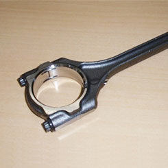 | Measuring a Connecting Rod’s Slit Width Using a Digital MicroscopeConnecting rods are required to be strong enough to withstand tens of millions of revolutions per minute, and the slit width is strictly controlled. With the DSX1000, the slit width that could not be clearly observed with a conventional microscope can be observed with high accuracy. |
|
|---|
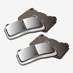 | Inspecting a Brake Pad’s Surface using a Digital MicroscopeA brake pad's surface impacts its performance, including braking force, heat stability, noise, and heat generation. Digital microscopes are used to check that the compounds used to create the brake pad are mixed properly. |
|
|---|
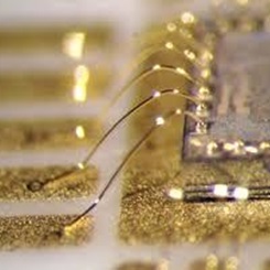 | Inspecting Bonding Wires Using a Digital MicroscopeDigital microscopes are effective tools for analyzing defects, such as wire breakage, wire pitch deviation, bonding peeling, and migration that can occur during the bonding process. |
|
|---|
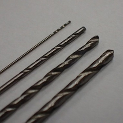 | Detecting Damage on a Drill Bit Edge Using a Digital MicroscopeDrill bits are widely used in industrial fields as a cutting tool. If the edge is damaged, inaccuracies may arise during hole positioning, or the drill may break. Conventional digital microscope is commonly used to perform drill inspection, but there are challenges. The DSX1000 offers advantages of detecting damage on a drill bit edge. |
|
|---|
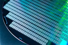
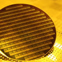 | Detecting Manufacturing Defects on Semiconductor Wafers Using a Digital MicroscopeSemiconductors are essential components in many electronic devices. Defects can be introduced into the circuit during manufacturing process, and visual inspection using a microscope is a preferred option to inspect defects. The DSX1000 simplifies semiconductor visual inspection. |
|
|---|
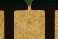
 | How a Digital Microscope’s Deep Focal Depth Enables the Complete Inspection of Connector PinsManufacturers use strict quality control measures to minimize failures of electrical connector pins, and microscopes play an essential role. The DSX1000 microscope’s objective lenses offer the depth of focus and resolution required to focus an entire connector pin at the same time, greatly simplifying and speeding up the inspection process. |
|
|---|
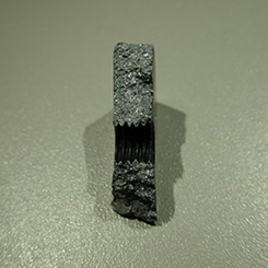 | Analyzing Fractured Metal Surfaces with a Digital MicroscopeFractography has become increasingly important as infrastructure continues to age and quality control issues cause problems. Optical or digital microscopes are essential fractography tools that are used to capture high-quality images for analysis. See details of advantages which DSX1000 can offer to analyze factured metal surfaces. |
|
|---|
 | Measuring the Volume of Integrated Circuit Chipping After the Dicing Process Using a Digital MicroscopeDuring the dicing process of integrated circuit (IC) manufacturing, amount of allowable roughness of wafer surface is carefully controlled. Amount of roughness is checked with a digital microscope, but the physical properties of IC chips can be challenging. The DSX1000 objective lenses offer high resolution at low magnification to reduce shading and flare, enabling inspectors to more easily see chipping during low-magnification observations. |
|
|---|
 | Inspecting Glass Fiber Peeling in a Printed Wiring Board’s Glass Epoxy Substrate—Clear Images Are Essential for Quality ControlInspection of resin peeling defects is critical as these defects can cause a completed PWB to have lower insulation and heat resistance, making them more susceptible to failure. PWBs are challenging to inspect with a microscope. The DSX1000 digital microscope has advanced telecentric optics and high-resolution objectives that offer an excellent depth of focus, which enable you to observe an etched PWB to investigate the cause of a defect. |
|
|---|
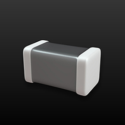 | Acquiring Clear Images and Accurate Dimension Measurements of a Laminated Ceramic Capacitor Using a Digital MicroscopeManufacturers measure the laminated ceramic capacitors’ dimensions and visually inspect them to look for cracks in the ceramic. Microscopes or digital microscopes is used to supplement the automated inspection system, but poses challenges. The DSX1000 offers multiple advantages to inspect capacitors. |
|
|---|
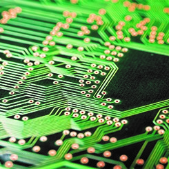 | Measuring the Circuit Shape of a Printed Wiring Board Using a Digital MicroscopeDuring the manufacturing process of PWBs, a microscopic inspection is necessary to analyze circuit shape precisely. There are multiple advantages of measuring circuit shape with the DSX1000. |
|
|---|
Automotive |
Electronics |
Metal Fabrication/Mold |
Chemical/Materials, Glass, and Ceramics |
Other |
