Optical Microscopes
Types of Optical (Incident Light) Microscopes
Optical microscopes are categorized on a structure basis according to the intended purpose. An upright microscope (left photo) which observes a specimen (object to be observed) from above is widely known as the most common type with a multitude of uses. An inverted microscope (right photo) which observes a specimen from beneath is used for observing the mineralogy and metallogy specimens, etc.

Microscope Basic Functions and Optical System Configuration
An optical microscope consists of the following two major basic functions.
- Creating a Magnified Image of a Specimen
- Illuminating a Specimen
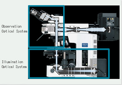
The function to create a magnified image of a specimen consists of three basic functions of "obtaining a clear, sharp image", "changing a magnification", and "bringing into focus". An optical system for implementing these functions is referred to as an observation optical system.
Meanwhile, the function to illuminate a specimen consists of three basic functions of "supplying light", "collecting light", and "changing light intensity". An optical system for implementing these functions is referred to as an illumination optical system. In other words, the observation optical system projects a sample (specimen) through an optical system and moreover leads a projection image to eyes or a pickup device such as CCD.
On the other hand, the illumination optical system effectively collects light emitted from the light source and leads the light to a specimen to illuminate it. The layout of observation and illumination optical systems in an optical microscope is as in the figure below for an upright microscope. Meanwhile, for an inverted microscope the layout relation between those optical systems is upside down at the center of a specimen with respect to an upright microscope.
Microscope Optical System Configuration

Principle of Optical Microscope (Compound Microscope)
An optical microscope creates a magnified image of an object specimen with an objective lens and magnifies the image further more with an eyepiece to allow the user to observe it by the naked eye. Assuming a specimen as AB in the following figure, primary image (magnified image) A'B' of inverted real image is created with an objective lens.
(ob). Next, arrange the eyepiece (oc) so that primary image A'B' is located closer to the eyepiece than the anterior focal point, then more enlarged erect virtual image A"B" is created. Put your naked eye in the eye (pupil) position on the eyepiece barrel to observe the enlarged image. In short, the last image to be observed is an inverted virtual image. As described above, this type of microscope which creates a magnified image by combining an objective lens making an inverted real image and an eyepiece making an erect virtual image is called a compound microscope. The observation optical system in an optical microscope is commonly standardized on this compound microscope. Meanwhile, such type of microscope that directly observes an inverted real image magnified with an objective lens is called a single microscope. A microscopic observation on a TV monitor, recently popularized increasingly, uses the way of directly capturing this inverted real image with a CCD camera, thereby being comprised of a simple microscope optical system.
Principle of Optical Microscope

Related Link
> Top of Digital Microscope page