Executive Summary
At the department of Conservation Studies at the University of Antwerp (Belgium), researcher Dr Olivier Schalm is employing the latest high-resolution light microscopy to reveal interesting new avenues of exploration within cultural heritage.
As an analytical technique, light microscopy provides unique insights across many areas of historical materials, from discovering an artefact's history to understanding the best course of action for its preservation in the future.
Unlike the majority of industrial samples, historical artefacts exhibit a high level of heterogeneity, and analysing the microstructure reveals information that otherwise remains hidden from the naked eye. Take the case of a paint layer detaching from its support, when inspected using light microscopy, the problem was found to originate from pitting corrosion of the underlying zinc plate. Identifying the particular species of oak forming a piece of furniture is even possible through analysing its cellular structure, providing clues regarding its geographical origin. In situations such as these, the types of materials and processes involved are much more evident at the microscopic level.
In addition to applied cultural heritage applications, light microscopy is also important for discovering novel conservation strategies. Only by truly understanding the process of degradation, one can develop and optimise techniques to control or slow down the process, or even restore the objects to a past state. The latter concept is a main focus of Dr Olivier Schalm's research at the Department of Conservation Studies.
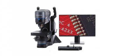
While the light microscope presents effective means of gathering information, if the investigation calls for greater resolution, researchers often turn to scanning electron microscopy (SEM). As technology has advanced however, the latest light microscopes such as the Olympus DSX510 are capable of reaching far higher resolutions than before, with magnifications of up to 4,000x now possible. In many cases, magnifying above this level is unnecessary, and with light microscopy not only retaining true-colour information but also utilising the latest digital imaging technologies, this approach can often present an attractive alternative to SEM. For cultural heritage research, the recent introduction of the Olympus DSX510 high-resolution light microscope in Dr Schalm's department has facilitated many studies: from advancing the understanding of glass and metallic degradation, to analysing damage on antique photographic plates.
Light microscopy in cultural heritage
Understanding the past
- Placing an artefact in its socio-cultural context – how/when/where was it created?
- Identifying, analysing and interpreting the component materials
- Understanding the process of degradation
Deciding the future
- Preventing degradation by manipulating the storage environment
- Choosing the best intervention for conservation or restoration
- Ongoing evaluation of protective measures
Re-thinking glass corrosion
Although glass begins life as a homogenous and transparent substance, over time it becomes gradually more opaque and irregularities begin to appear. Since Sir David Brewster's efforts in the 19th century, this process was thought to be fully understood. However, looking at glass artefacts in more detail has shown us that this process is perhaps more complex than previously thought, and Figure 1 illustrates an historic glass sample exhibiting an unusual characteristic.
Running right through the structure are lamellae formations, and it remains unknown as to how they form, including reasons for the difference between the black and white rings, and the basis for the different results generated from brightfield and darkfield illumination. Detailed analysis over a large area of the sample is crucial for these ongoing studies, and image stitching is performed using the DSX510, enabling the glass to be viewed at a magnification of 555x over a field of view of 1.4
mm2. Moreover, like most antiques, the surface of old window glass samples is never perfectly flat, and in the past, generating such sharp images at high magnification was not possible with light microscopy. By creating a z-stack with the Extended Focal Image (EFI) function of the DSX510, it is now possible to acquire sharp images of antique samples, enabling detailed analysis and facilitating our understanding of how these irregularities form.
Employing different illumination techniques is also useful for visualising different forms of glass corrosion, such as the formation of manganese-rich inclusions. The glass artefact shown in Figure 2 has developed these inclusions over the last 200 years or so, appearing in dendritic formations. While brightfield observation displays the inclusions on the surface due to a higher refractive index, visualisation using darkfield illumination reveals them beneath the surface. This unexpected
finding indicates an intricate three-dimensional structure, without the need for complex tomography experiments. Since this form of manganese is non-mobile, it is suspected that a water-soluble form first travels towards certain regions where an redox reaction takes place, and research aims to elucidate this process further.
Inspecting the degradation of glass.
| Surface of a window glass fragment - visualising lamellae in unprecedented detail was achieved with darkfield illumination at 555x magnification over a field of view of 1.4 mm2, employing image stitching and Extended Focal Imaging capabilities to form a large, fully-focused image. |
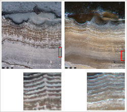 Figure 1.B : Brightfield(left) and Darkfield(right) | Cross section of the sample fragment shows the lamellae running internally, differentially illuminated with brightfield and darkfield at 1,000x magnification, providing further insights into these structures. |
Investigating the formation of mineral inclusions in glass.
Manganese inclusions formed during the last 200-yearsare visualised with brightfield and darkfield illumination at 100x magnification. Employing the Extended Focal Imaging function creates a fully focused image, and combined with darkfield visualisation shows how the dendritic inclusions are not only superficial, instead running beneath the surface.
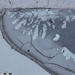 Figure 2.A : Brightfield | 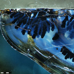 Figure 2.B : Darkfield |
Images acquired using the Olympus DSX510; courtesy of Dr Olivier Schalm, University of Antwerp.
The beginnings of photography: Daguerreotype conservation
Introduced in 1839, the daguerreotype was the first mainstream photographic process. An image is formed due to the scattering of light by nanoparticles of silver and mercury lying upon a polished silver surface, meaning that both scratches and tarnishing can easily obscure the image – an example of this is shown in Figure 3. Through understanding the process of silver corrosion it becomes possible to optimise cleaning treatments and preserve these photographic plates in the future, but the damage must first be characterised.
One important aspect of analysing the damage of antiques is knowing where a feature sits within the context of the whole piece, as microstructures can differ significantly across the artefact. Every point on such an object is unique and this link is vital, and should be retained when investigating the sample at higher magnifications. Moving gradually towards a higher magnification with light microscopy, while at the same time being able to physically see where the light beam hits the artefact helps maintain this link. Navigation is also often achieved using cues from specific features, for example locating a scratch using the feature of the eye as a point of reference (Figure 3B). When comparing brightfield visualisation at 555x magnification with conventional secondary electron SEM imaging, the image of the eye is only visible with light microscopy. In order to observe a similar feature in a SEM, backscattered electron imaging (where contrast is based on a material's atomic number) needs to be used, which is a more expensive technique.
Profiling daguerreotype damage.
The silver plate of this daguerreotype (A) was analysed in more detail, with darkfield visualisation (positive image) creating a more natural image than in brightfield (negative image) (B). When compared with backscattered scanning electron microscopy (BS-SEM) to profile the location of a scratch at 555x magnification, light microscopy using the Olympus DSX510 delivered the contrast necessary to visualise the eye - a navigation feature (C).
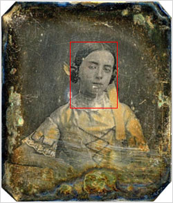 Figure 3.A |
|
Images courtesy of Dr Olivier Schalm and Eva Grieten, University of Antwerp.
Reversing corrosion
With modern conservation techniques, it is now possible not only to stop corrosion, but in some cases, actually reverse the oxidation state. This is achieved through atmospheric plasma afterglow treatments, and the efforts of Dr Schalm's research group in improving this technique are part of the European PANNA project ("Plasma and nano for new-age soft conservation"). Running over three years, the project focuses on new techniques for cleaning and protection of cultural heritage assets.
Hydrogen-based plasma creates a reactive species able to reduce the black tarnish of silver sulphide generated by corrosion, thereby restoring the distinctive glossy silver surface. Whilst pure silver can be reduced in a matter of seconds, copper is impervious to current plasma treatments, and introducing even the smallest amount of copper complicates the process. Unfortunately, historical silver mostly contains a small amount of copper (e.g., sterling silver is an alloy typically used for jewellery and consists of 92.5 w% Ag and 7.5 w% Cu) and as yet, the basis for this selectivity in the plasma chemical reactions remains unknown. A greater understanding is not only vital for improving the application of plasma treatment towards different materials, but also in avoiding unexpected damage being caused in the long-term.
By immersing a pure copper plate in an aerated sodium sulphide solution for 30 minutes, and inspecting the subsequent formation of copper sulphide compounds with high-resolution light microscopy, it was possible to analyse copper corrosion more in depth. Although SEM generates highly detailed images, these lack colour information, and high-resolution light microscopy has revealed new insights – with Figure 4 showing particles of a corrosion product forming preferentially at the metallic grain border. As a colour rather than morphological feature, these zones of green corrosion were previously missed using SEM, and this discovery was only possible with true colour information.
Tracking the progress of sterling silver corrosion from sodium sulphide exposure more closely has also yielded unexpected information, using light microscopy alongside gloss measurement (which indicates the smoothness of a surface) to analyse the ongoing effect of sodium sulphide (Figure 5). From the sequence of images captured over an hour at 2,000x magnification (Figure 5A), it can be seen that islands forming around the copper inclusions coincide with an associated drop in gloss. What is more surprising, however, is the subsequent increase in gloss occurring as these islands merge; thought to derive from this newly flattened and reflective surface. This is the point at which we see the silver begin to corrode, likely owing to the depletion of all copper at the surface, and by 60 minutes the surface has completely transformed. It was therefore a novelty to observe how exactly the copper dominates the corrosion process of sterling silver.
With high-resolution light microscopy allowing researchers to track the process of corrosion in unprecedented detail, it has become evident that the chemical reactions involved are more complex than first envisaged a couple of years ago. A particular requirement of such an experiment is the ability to place the sample back under the microscope at each interval, for immediate visualisation of exactly the same position – which would be much more cumbersome with SEM.
Visualising copper corrosion.
Copper was sulphidised in an areated Na2S solution and the surface of the metal coupon visualised using light microscopy after 30 minutes: at magnifications of: 150x (A), 2,000x (B) and 4,000x (C). Retaining true colour information at such high resolution revealed a green corrosion at the metallic grain border, which was missed during the inspection with SEM .

Figure 4.A : 150x Figure 4.B: 2,000x Figure 4.C: 4,000x
Visualizing the corrosion process of sterling silver.
Sterling silver immersed in Na2S was analysed with light microscopy at 2,000x using brightfield observation at different time intervals at exactly the same position as a function of exposure time (A). Copper inclusions within the silver matrix corrode first forming islands that are larger than the original inclusion. After 7 minutes, the copper sulphide islands merge, and silver corrosion begins (grey). (B) Gloss drops as surface becomes rougher from copper islands, but rises unexpectedly as they merge to form a flat, reflective surface. This once again drops with silver corrosion.
0 min.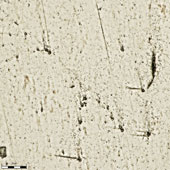 | 1 min.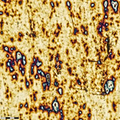 | 3 min.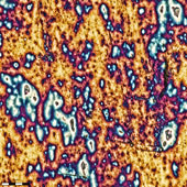 | 5 min.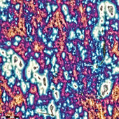 |
7 min.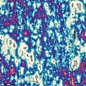 | 15 min.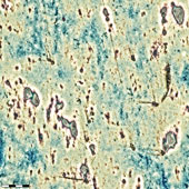 | 30 min.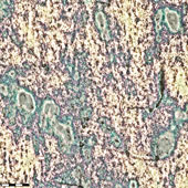 | 60 min. |
| Figure 5.A: Time (mins): | |||
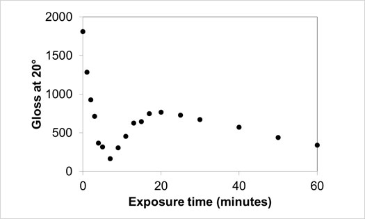
Figure 5.B: Gloss indicates surface topography
Images acquired with the Olympus DSX510;courtesy of Dr Olivier Schalm and Patrick Storme, University of Antwerp.
Summary
High-resolution light microscopy presents an insightful tool for cultural heritage investigations, allowing researchers to characterise damage and probe the subtle and complex changes taking place during the process of corrosion. Affording several major advantages, this approach has enabled Dr Schalm’s research group to easily extract new information from a range of historical artefacts.
Although it can sometimes be the case that studies call for complementary methods, for example with a higher magnification or information on chemical composition, high-resolution light microscopy presents a quick and efficient first-line analysis, which can be complemented with additional information as necessary.
For both glass and metallic artefacts, the information revealed within these studies has in some cases been rather unexpected, raising further questions relating to the mechanisms underlying such processes. There is still a way to go before our understanding of material corrosion is complete, but moving into the future it is clear that the latest high-resolution light microscopy will play an important role, rapidly advancing our knowledge of the past.
Author information
Dr Olivier Schalm is a lecturer and researcher at the Department of Conservation Studies at the University of Antwerp (Belgium).
Markus Fabich is Product Manager for Materials Science Microscopy at Olympus SE & CO. KG (Hamburg, Germany).


