BX61
Discontinued Products
Краткий обзор
Olympus BX61 microscopes were developed based on cutting edge optical and imaging technologies to address various materials science applications.
Olympus Is Dedicated to Making Microscope System Solutions to Support Your Work on All Levels
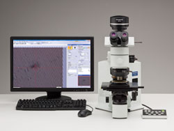 BX61TRF/BX-RLAA/U-AFA2M/DP Series | The BX61 is specially designed to work with the Olympus laser based autofocus unit. With precise focus and active tracking, users can speed up routine inspections. Additional settings such as illumination level, lens selection and aperture setting can be set using push buttons on the microscope frame, a keypad or via the PC. A variety of motorized modules including nosepieces and illuminators are available to provide you with full flexibility in configuring your system. |
Excellent Image Clarity and Superb Resolution
Choose from a Range of Objective Lenses to Customize Your System
Olympus offers a wide variety of objective lenses to suit every observation technique. You can select the right lens for your application from our family of over 150 objective lenses. Color fidelity is important for accurate, efficient inspection. The UIS2 objective lens series yields natural color reproduction by combining carefully selected high-transmittance glass and advanced coating technology.
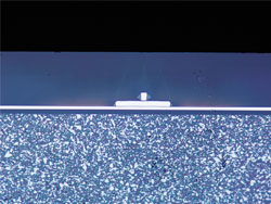
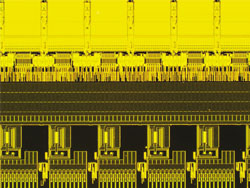
Example Observation Images
 Magnetic Head (Brightfield) | 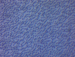 DVD (Brightfield) |
 Wafer (Fluorescence) |  Color Filter (Transmitted) |
| Diverse Lineup Allows Selections According to the Purpose | |
| MPLAPON | Plan Apochromat Objective Lens Series |
| MPLFLN | Semi Apochromat Objective Lens Series for Brightfield |
| MPLFLN-BD | Semi Apochromat Objective Lens Series for Brightfield and Darkfield |
| MPLFLN-BDP | Semi Apochromat Objective Lens Series for Brightfield, Darkfield adn Phase Contrast |
| LMPLFLN | Long WD Semi Apochromat Objective Lens Series for Brightfield |
| LMPLFLN-BD | Long WD Semi Apochromat Objective Lens Series for Brightfield and Darkfield |
| MPLN | Plan Achromat Objective Lens Series for Brightfield |
| MPLN-BD | Plan Achromat Objective Lens Series for both Brightfield and Darkfield |
| SLMPLN | Super Long WD Plan Achromat Objective Lens Series |
| LCPLFLN-LCD | Long WD Semi Apocromatic Objective Lens Series for LCD |
| LMPLN-IR/LCPLN-IR | Long WD Plan Achomatic Objective Lens Series for Near Infrared light |
> Click here for details about UIS2 objective lenses
Software Solutions
Built to Easily Work with Digital Cameras and Image Analysis Software
Our full line of digital cameras provide high resolution viewing and fast image transfer while our advanced OLYMPUS Image Analysis Software empowers users and provides all the tools required for today's most complex metallurgical requirements. Choose from Extended Measurements,Standard Metallography and Advanced Metallography application specific modules (over a dozen application specific routines) and automatically populate data and create reports in compliance with the most popular ASTM and ISO specifications, all with just a few clicks of the mouse.
Характеристики
| Optical system | UIS2 optical system (infinity-corrected) |
|---|---|
| Microscope frame > Illumination | Reflected/transmitted |
| Microscope frame > Illumination |
External 12 V 100 W light source
Light preset switch LED voltage indicator Reflected/transmitted changeover switch |
| Microscope frame > Focus |
Motorized focusing
Stroke 25 mm Minimum graduation 0.01 μm |
| Microscope frame > Max. specimen height | 25 mm (w/o spacer) |
|
Observation tubes > Widefield
(F.N. 22) |
Inverted: binocular, trinocular, tilting binocular
Erect: trinocular, tilting binocular |
|
Observation tubes > Super widefield
(F.N. 26.5) |
Inverted: trinocular
Erect: trinocular, tilting trinocular |
| Reflected light illumination > BF etc. |
BX-RLAA
Motorized BF/DF changeover Motorized AS |
|
Reflected light illumination > Reflected
fluorescence |
BX-RFAA
Motorized 6 position turret Built-in motorized shutter With FS, AS |
| Transmitted light |
100W halogen
Abbe/long working distance condensers Built-in transmitted light filters (LBD, ND25, ND6) |
|
Revolving
nosepieces > For BF | Motorized sextuple |
|
Revolving
nosepieces > For BF/DF | Motorized quintuple, motorized sextuple, centering quintuple |
| Stages |
Coaxial left(right) handle stage: 76 (X) x 52 (Y) mm, with torque adjustment
Large-size coaxial left (right) handle stage: 10 0(X) x 105 (Y) mm, with lock mechanism in Y axis |
| Dimensions |
Approx.
318 (W) x 602 (D) x 541 (H) mm |
| Weight |
Approx. 25.5 kg
(Microscope frame 11.4 kg) |
Фотогалерея применения
BX61 Gets You the Image You Want
Darkfield
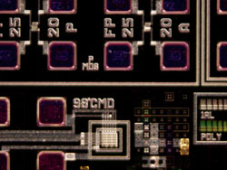 Surface mounting board | Darkfield lets you observe scattered or diffracted light from the specimen. The light from the lamp travels through ring-form illumination optics in the illuminator and is focused on the specimen. The light from the specimen is reflected only by imperfections in the z axis. The user can identify the existence of even a minute scratch or flaw down to the 8 nm level--smaller than the resolving power limit of an optical microscope. Darkfield is ideal for detecting minute scratches or flaws on a specimen and examining mirror surface specimens, including wafer. |
Polarized Light
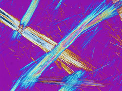 Asbestos | This microscopic observation technique utilizes polarized light generated by a set of filters (analyzer and polarizer). The characteristics of the sample directly affect the intensity of the light reflected through the system. It is suitable for metallurgical structures (i.e., growth pattern of graphite on nodular casting iron), minerals, and LCDs and semiconductor materials. |
Differential Interference Contrast (DIC)
 Magnetic head | DIC is a microscopic observation technique in which the height difference of a specimen not detectable with brightfield becomes a relief-like or three-dimensional image with improved contrast. This technique, based on polarized light, can be fitted to your needs with a choice of three specially designed prisms. It is ideal for examining specimens with very minute height differences, including metallurgical structure, minerals, magnetic heads, and hard-disk media and polished wafer surfaces. |
Fluorescence
Particle on semiconductor wafer | This technique is used for specimens that fluoresce (emit light of a different wavelength) when illuminated with a specially designed filter module that can be tailored to your application. It is suitable for inspection of contamination on semiconductor wafers, photo-resist residues, and detection of cracks through the use of fluorescent dye. An optional apochromatic lamp house collector lens system can be added to compensate for chromatic aberrations from visible light to near-infrared light. |
Transmitted Light Observation
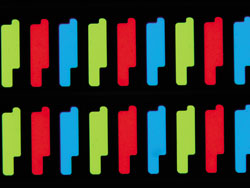 LCD color filter | For transparent samples such as LCDs, plastics and glass materials, true transmitted light observation is available by using a variety of transmitted light condensers. You can examine your samples in brightfield, darkfield, DIC, and polarized imaging in transmitted light, all in one convenient system. |